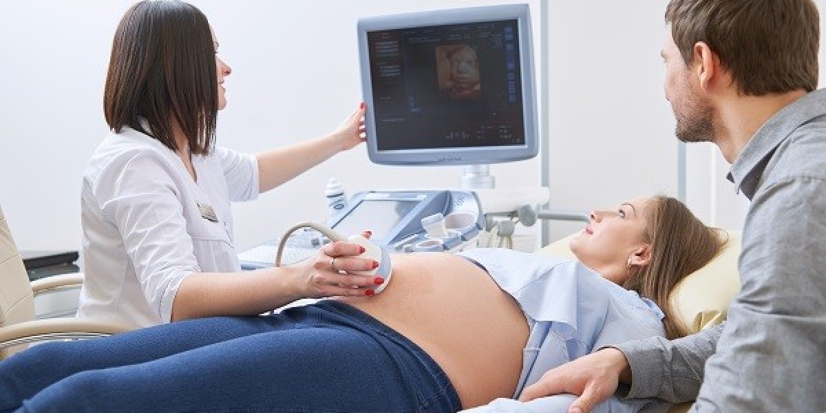
Sonography, or ultrasound imaging, is a crucial diagnostic tool that utilizes high-frequency sound waves to create images of the body's internal structures. This non-invasive technique is widely used in various medical fields, including obstetrics, cardiology, and emergency medicine, due to its safety and effectiveness. At CT Nursing Home in Dhanori, patients benefit from advanced sonography services that leverage state-of-the-art technology and skilled professionals. The facility is dedicated to providing high-quality care and accurate diagnoses, making it a premier destination for sonography services in Dhanori.
How Sonography Works:
Basic Principles of Sonography
Sonography operates on the principle of sound wave reflection. An ultrasound machine consists of a transducer that emits high-frequency sound waves (typically between 1 MHz and 18 MHz) into the body. When these sound waves encounter different tissues, they are reflected to the transducer at varying degrees based on the density and composition of the tissues.
Sound Wave Emission: The transducer sends out sound pulses into the body.
Echo Reception: The sound waves bounce back from tissues and are received by the same transducer.
Image Formation: The ultrasound machine processes these echoes to create visual images of internal structures.
The resulting images, known as sonograms or ultrasonograms, provide valuable information about the size, shape, and consistency of organs and tissues.
Types of Ultrasound Modes:
Different modes of ultrasound are utilized depending on the diagnostic needs:
A-mode (Amplitude Mode): This one-dimensional mode displays the amplitude of received echoes as a function of time. It is primarily used for measuring distances.
B-mode (Brightness Mode): The most commonly used mode, B-mode produces two-dimensional images by displaying brightness levels corresponding to echo amplitudes. This allows for detailed visualization of internal structures.
M-mode (Motion Mode): Used mainly in cardiology, M-mode captures motion over time, providing information about moving structures such as heart valves.
Doppler Ultrasound: This technique measures blood flow by assessing changes in frequency (Doppler effect) as sound waves bounce off moving red blood cells.
Safety and Efficacy
Sonography is considered a safe procedure with no known harmful effects when performed by trained professionals. Unlike X-rays or CT scans, ultrasound does not use ionizing radiation, making it suitable for a wide range of patients, including pregnant women.
Technology Used in Ultrasound Machines at CT Nursing Home:
CT Nursing Home employs state-of-the-art ultrasound technology to ensure high-quality imaging and accurate diagnostics. The following sections detail some key technological features integrated into their ultrasound machines.
Advanced Transducer Technology
Transducers are crucial components of ultrasound machines. They convert electrical energy into sound waves and vice versa. At CT Nursing Home, various types of transducers are utilized:
Linear Array Transducers: Ideal for superficial structures such as veins and arteries.
Convex Array Transducers: Used for abdominal imaging due to their wider field of view.
Endocavitary Transducers: Employed for specialized examinations such as transvaginal or transrectal ultrasounds.
ZONE Sonography Technology (ZST):
CT Nursing Home utilizes advanced ZONE Sonography Technology (ZST), which revolutionizes traditional ultrasound imaging techniques. ZST allows for enhanced image formation through:
Dynamic Pixel Focusing: This technology improves image quality by utilizing data from multiple overlapping zones to focus sound waves more effectively at various depths.
Automated Sound Speed Compensation (SSC): SSC recalibrates the speed of sound based on tissue characteristics during imaging, enhancing spatial resolution and contrast in images.
These innovations lead to clearer images that facilitate accurate diagnoses.
Image Processing Software:
The ultrasound machines at CT Nursing Home are equipped with sophisticated image processing software that enhances image quality in real-time. Key features include:
CrossXBeam™ Technology: This spatial compounding technique captures multiple images from different angles and combines them into a single high-resolution image. It reduces noise and improves anatomical clarity.
Automated Image Optimization: The software automatically adjusts settings based on patient anatomy and examination type, ensuring optimal image quality without requiring manual adjustments from sonographers.
Integration with Electronic Health Records (EHR)
To streamline patient care, CT Nursing Home integrates its ultrasound systems with electronic health records (EHR). This allows for:
Immediate Access to Results: Physicians can view ultrasound results promptly within patient records.
Enhanced Communication: Improved data sharing between departments facilitates comprehensive patient management.
Benefits of Sonography Services in Dhanori at CT Nursing Home
CT Nursing Home has positioned itself as a leader in providing sonography services in Dhanori through its commitment to quality care and advanced technology. The facility offers a wide range of ultrasound services tailored to meet various diagnostic needs:
Obstetric Ultrasound: Essential for monitoring fetal development during pregnancy.
Abdominal Ultrasound: Used to assess organs such as the liver, kidneys, and gallbladder.
Cardiac Ultrasound (Echocardiography): Evaluates heart function and structure.
Doppler Studies: Assesses blood flow in vessels.
Musculoskeletal Ultrasound: Diagnoses conditions related to muscles and joints.
Conclusion:
In conclusion, sonography is an essential component of modern medical diagnostics, offering valuable insights into the human body without invasive procedures. At CT Nursing Home in Dhanori, the integration of advanced ultrasound technology and experienced professionals ensures that patients receive top-notch care tailored to their specific needs. The facility employs cutting-edge features such as advanced transducers and sophisticated image processing software to enhance image quality and diagnostic accuracy. Furthermore, the commitment to patient comfort and thorough follow-up by skilled staff reinforces CT Nursing Home's reputation as a leader in providing sonography services in Dhanori. As healthcare continues to evolve, the role of sonography will expand, further contributing to early diagnosis and effective treatment of various medical conditions.
